The Hidden Harms of Modern Lighting and the Fight to Bring Back Incandescent Bulbs
**Story at a Glance: •Sunlight is one of the most important things for our health (e.g., it halves our risk of dying or getting cancer), yet all we ever hear is how important it is to avoid it.**
**•While the importance of vitamin D is generally recognized, many of the other critical functions of light within plants, animals and human beings remain almost completely unknown, and hence are the focus of this article.**
**•One of the defining characteristics of modern life is continual exposure to unnatural light (particularly from energy efficient light bulbs). This is viewed as relatively benign, but decades of forgotten research (and a growing body of new research) show it is a root cause of a myriad of inexplicable physical and behavioral illnesses.**
**•Cells communicate with each other through faint emissions of ultraviolet light. This was once one of the most active fields in biology (yielding over 700 papers, 11 Nobel nominations, and a blood test which detected cancer with over 95% accuracy) before it was abandoned for political reasons and forgotten.**
**•The eyes and the blood conduct light throughout the body. When this process is disrupted (e.g., by glasses with lenses that filter out critical parts of the spectrum), a variety of significant illnesses can develop.**
**•In 2022, the federal government effectively banned the incandescent bulb because most of its output is “wasted” infrared rather than visible light, despite our bodies using that infrared for a myriad of essential processes. Senate Energy Chairman Mike Lee introduced a bill to bring those bulbs back, which yesterday received its first hearing, and with public support, can free us from the unhealthy bulbs that have been forced upon us.**
Over the years, I have become convinced sunlight is one of the most essential nutrients for the body. For example, as human beings moved further and further north from Africa and had increasingly lower sun exposure, their skin became progressively lighter, something which argues the human body needs light and that the whitening of human skin in the higher latitudes was a protecting mechanism designed to ensure enough of the remaining sunlight could get inside the body. _Note: an explanation of how the darker parts of the skin prevent UV from entering the body can be found here._
Unfortunately (outside of very specific patentable applications), since no one has a financial interest in promoting sunlight it hence has no lobbyists, almost everything we hear about instead focuses on why it’s terrible for us (as sunlight prevents many diseases other industries profit from). For example:
•As discussed in a previous article, dermatology was one of the least desirable professions in medicine. They then transformed themselves into one of the most sought after ones by rebranding themselves as cancer fighters and convincing everyone to get regular skin cancer exams where any potential cancer could be quickly removed, often for thousands of dollars apiece, quickly making dermatology into one of the highest paying specialties. A centerpiece of this rebranding was making benign cancers be labeled as deadly ones and claiming sunlight caused skin cancer (which while true, omitted to mention that that **a lack of sunlight** is what causes fatal skin cancers). Because of this, a lot of dermatology morphed into scaring people senseless about the sun, and despite billions being spent each year on skin cancer, the total death rate from skin cancer remains almost entirely unchanged (instead we simply diagnose and “treat” far more of it). _Note: in this article, I will attempt to explain why unnatural light causes skin cancer._
•While many factors contribute to cancer, the focus is often on sunlight and smoking as the primary culprits. I believe this emphasis explains why cancer rates continue to rise, as the true causes of cancer remain underexplored. Industries with vested interests lobby to keep these causes off-limits, diverting attention from potentially significant contributors to cancer.
In this article, I will present some of the forgotten knowledge on the incredible importance of light.
Prior to dermatology’s disastrous war on the sun, the value of sunlight was widely recognized in medicine. For example, in the early 1900s, heliotherapy (sunbathing) was used with great success for treating many (otherwise incurable) conditions, such as the 1918 influenza, tuberculosis, and many other diseases.
The existing data, in turn, shows that sunlight provides an immense degree of benefit. For example:
•Sunlight is critical for mental health. This is most well appreciated with depression (e.g., seasonal affective disorder). Sadly, many workers (especially night shift workers such as those in hospitals) do not get that essential light exposure and suffer immensely. For example, consider this study of Chinese operating room nurses which found their mental health was significantly worse than the general population and that this decline was correlated to their lack of sunlight exposure (which I would argue does not benefit the patients they care for).
_Note: I really got this point once, where after a long period of hospital night shifts under fluorescent lights, noticed I was becoming clinically depressed (which has never otherwise happened to me and led to another physician I was close to offering to prescribe antidepressants). I decided to do an experiment and stuck with it for a few more days, then went home and bathed under an Ott full spectrum bulb I’d been able to obtain, at which point I almost instantly felt better._
•Many different studies have found sunlight exposure dramatically reduces one's risk of cancer (e.g., this large study found high solar UVB exposure halved one’s risk of breast and prostate cancer).
_•_An excellent 20-year study of 29,518 women found that avoiding the sun made one 60% more likely to die (a 130% difference compared to those who had the most significant amount of sun exposure), and the most considerable benefit from regular sunlight exposure was reducing one’s risk of dying from heart disease. _Note: that study also found a variety of other common diseases were much less likely to affect those with adequate sun exposure._
_•_In a 2024 analysis of the UK Biobank, both measures of UV exposure that were examined (sunbed use and how much solar radiation reached the participant’s home) were associated with a lower risk of dying from any cause, from cardiovascular disease and from cancer.Likewise, when88,905 Biobank participants wore light sensors for a week, those with brighter days and those with darker nights were each less likely to die over the following 8 years.
•Normal sunlight is critical for facilitating the circadian rhythm our body’s depend upon to rest and repair themselves. In turn, a major cause of the modern insomnia epidemic (and the profound health consequences it entails—which are discussed further here) are artificial light exposures, while one of the most useful treatments for it is to simply start your day with a full sunlight exposure.
Typically, sunlight's benefits are thought to be due to producing vitamin D (a critical nutrient) within the body. However, I have long suspected that many of the benefits of vitamin D are not due to the vitamin itself but rather that its elevation serves as an indicator that the body is having regular sunlight exposure and hence is experiencing the myriad of benefits sunlight provides. For example, when 24 healthy volunteers were exposed to UVA (which does not produce vitamin D) at a dose equivalent to about 30 minutes of summer sun, their arteries dilated and their blood pressure dropped, as nitric oxide stored in the skin was released into the circulation.The dermatologist behind that work, has since argued (e.g., in this review) his specialty’s position on sunlight needs to be rethought. Similarly, a meta-analysis found that spending an hour or more a day in the summer sun was associated with a 16% lower risk of breast cancer, whereas simply living somewhere with more ambient UV was not.
_Note: that being said, vitamin D supplementation can often be very beneficial. However, in almost all cases, elevating your vitamin D levels through appropriate sunlight exposure provides significantly greater benefits than supplementation._
Waves, by definition have a few fundamental properties:
•They require a medium to travel through. For example, the medium for waves at the beach is the ocean, whereas the medium for sound is the air.
•They travel at specific speeds.
•They have a specific wavelength and frequency (which are inversely related to each other).
•Their strength is determined by how many waves elapse in a second (a higher frequency correlates to a higher total energy) and how greatly each individual wave disrupts its medium (e.g., a small wave at the beach has much less energy than a tsunami as does a loud sound compared to a quiet one).
By far, the most common wave in our lives is electromagnetic radiation, which is defined as a wave that travels at the speed of light and, unlike all other waves, does not have a medium it travels through (which is why EMR can travel through space and many objects). “Light,” for instance, is one type of EMR.Since there is an inverse relationship between a wave’s wavelength and frequency, a variety of different types of EMR exist depending upon their wavelength. For example, visible light comprises the EMR with a wavelength between 380nm-700nm, as this is the EMR the eye can “see.” At the same time, depending on the wavelength of the EMR, its properties (e.g., color) vary immensely.
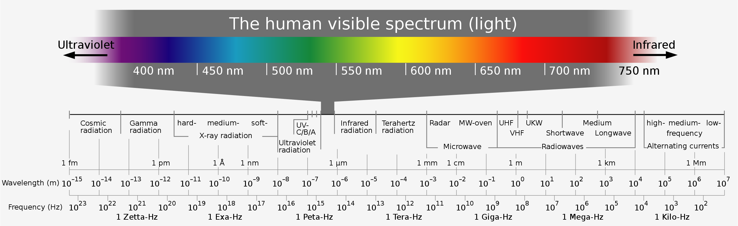
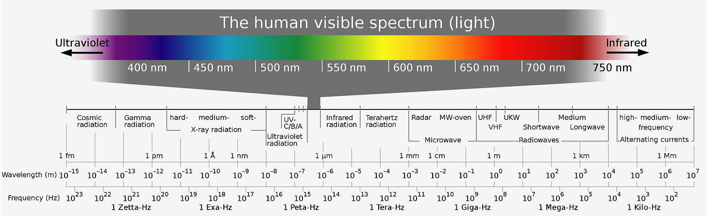
Likewise, many other types of EMR also exist (e.g., Gamma rays, x-rays, ultraviolet, visible light, infrared, microwaves, and radiowaves) which each have different properties. However, despite EMR being one of the fundamental constructs of our reality, much of its biological significance is still not recognized. For example, I feel some of the biggest misunderstandings about EMR are as follows:
•Natural light tends to have a fairly wide range of frequencies present within it (e.g., this is why sunlight splits into a rainbow once it passes through a prism). In contrast, artificial light tends to have a much narrower range of frequencies present within it which differs greatly from what biology evolved to handle.
•This is easiest to see by comparing bulbs. A standard white LED emits a sharp spike of blue at around 450nm, a broader hump of yellow, and almost nothing past 700nm. An incandescent bulb, like the sun, emits a smooth curve that keeps rising through the red and continues past 2000nm, so most of what it produces is infrared we cannot see.1_Note: since the government grades bulbs on visible light per watt, that infrared was scored as “waste,” which is how the incandescent bulb came to be banned._
•The label “ultraviolet” is given to the EMR that lies between visible light and x-rays. This implies all UV is the same, when in reality, each type of UV (UV-A, UV-B, and UV-C behave very differently) and are present in very different amounts (e.g., the atmosphere blocks most UV-C from reaching the surface of the Earth).
•Infrared penetrates much deeper into the body than UV (5-40mm vs. 0.02-0.15mm) and in 2025, researchers at University College London were able to measure the infrared in sunlight after it had passed through the human body and found that it created systemic effects.Additionally, standard glass (for the most part) blocks UV from traveling through it.
•Conventional science typically describes the adverse effects of EMR as being a property of how much energy the EMR has and if that energy is sufficient to break molecular bonds (termed ionizing energy). For example, gamma rays (which are released by nuclear weapons) are so dangerous because their ionization energy is so powerful they shred the molecular structures of the body (e.g., DNA) apart. Conversely, microwaves (which are instead on the low energy end of the electromagnetic spectrum) are deemed to not be dangerous because they contain too little energy to ionize molecular bonds.
•The previous point is not entirely correct because while EMR typically passes through whatever it contacts, if it instead “matches” that medium, it will store inside it and have a dramatically greater effect on it (e.g., this describes the concept of a resonance frequency). This is important because a wide number of critical living structures (e.g., mitochondria) evolved to have a resonance for a specific type of EMR and then respond to them while everything around them is largely unaffected by that specific type of EMR. Unfortunately, this aspect of science has been largely glossed over by our scientific apparatus, something I believe is due to the fact biophysics opens the doors to creating a variety of un-patentable medical therapies, and because it implicates a lot of the EMR we are now saturated in (e.g., the microwaves cell phones bluetooth and Wifi use) as a cause of poor health.
For example, radar works by sending out a large pulse of microwave energy in each direction (through the unit spinning around), and then using the reflections it receives from any radar pulse into the atmosphere to indicate the presence of an object being there. I personally think radar is far from benign as I’ve read numerous stories of soldiers stationed next to radar units (where the microwaves are exponentially stronger) becoming severely ill, I know many EMF sensitive people can tell if far away radar units are firing, and I personally can feel something when I am at an airport each time a radar sweeps by. While that seems abstract, I recently saw a brief video by John Ott that perfectly demonstrates the reality of this phenomenon:
_Note: in another article I wrote on this critical subject I highlighted how it underlies many of the therapies now used in regenerative medicine._
A fundamental principle within biophysics is that cells emit very faint photons (predominantly within the ultraviolet spectrum) they use to control growth and communicate with other cells and that when biophoton transmissions go awry, disease results (leading to abnormal photon emissions from tissue being seen in many disease states). For example, cancers have abnormal biophoton emissions, and (when studied) carcinogenic substances significantly disrupt the wavelength of these photons, whereas similar compounds that do not disrupt those biophotons are not carcinogenic (an observation which led Fritz-Albert Popp to create the discipline of biophotonics).
One of the most interesting observations made within biophotonics was that the cytopathic changes caused in a cell by viral infections or toxin exposures could be “transferred” to another cell in the immediate vicinity when the cells had no physical connection but were optically connected through a UV transmitting window, while conversely (as this Russian study shows), cells injured by radiation could be healed by being placed by healthy ones.
_Note: when cells are irradiated, nearby unirradiated cells can also show radiation-like injury (known as the bystander effect). Recently, evidence has accumulated that ultra-weak biophoton emission contributes to the bystander effect.1,2,3_
Likewise, in health, biophoton emissions cycle with the circadian rhythm (e.g., when a Japanese team imaged the light coming off the human body, it peaked in the late afternoon and stayed low overnight, even in volunteers kept awake under constant light1), whereas in chronic illness, Robert Naviaux (the researcher who pioneered studying the cell danger response) mapped each stage of the cell danger response to a characteristic level of emission the body becomes stuck at.1
Alexander Gurwitsch was one of the earliest researchers to study this. In 1923, he discovered that living cells (of both animals and plants) gave off extremely faint emissions which triggered the cells around them to leave their lag phase, enter mitosis and divide, leading him to name it mitogenic radiation [MGR]. After realizing that ordinary glass but not quartz glass blocked it, he concluded that MGR was a type of ultraviolet light (as this is how UV behaves), and much later, it was confirmed to be exactly that (with a wavelength between 190-350 nm).
_Note: MGR is very faint (making it difficult to detect), and its emission from biological systems typically requires the system to be illuminated with light (which makes the faint mitogenic emissions much more difficult to spot). Eventually, roughly a decade after Gurwitsch’s death, researchers were able to capture this radiation with sensitive photomultiplier devices._
After studying it in detail, Gurwitsch realized that MGR required a very specific dosing and pattern (e.g., through being pulsed) to achieve its optimal effects, and that too much quickly became counterproductive (e.g., light UV exposure stimulated the growth of yeasts, while stronger UV exposure killed them). For example, light interrupted 100-800 times a second produced the effect in 13 seconds (versus 6-8 minutes for continuous light), while yeast responded to the UV from living tissue at intensities orders of magnitude lower than they did to UV from a lamp, and responded far better to a lamp emitting a mix of wavelengths than to one emitting a single spectral line.1
As this was very challenging to do artificially, the value of MGR was primarily in what it facilitated the diagnosis of (e.g., blood typically emitted MGR, but if someone had cancer it did not, and as a result, with a high degree of accuracy, the hospital he worked at was able to determine if someone had a cancer purely by assessing if MGR had disappeared from their blood). This was eventually traced to a peptide which appeared in the blood of cancer patients and quenched its UV emission (termed the “cancer quencher”), whereas in the other states where blood stopped radiating (e.g., fatigue, starvation, old age, diabetes or sepsis), this was instead due to metabolic byproducts which absorbed the UV, a difference the test could distinguish. In animals, the quencher appeared long before a tumor could be felt (e.g., in rats, 9-12 days after tumor cells were implanted versus 28-32 days) and disappeared within days of the tumor being successfully removed (while its persistence predicted a recurrence). Likewise, in the clinics which used it to screen newly admitted patients, both its sensitivity and specificity exceeded 95%, roughly thirty years before the first conventional blood tumor marker (alpha-fetoprotein) was discovered.1
Additionally, Gurwitsch and those who followed him (many of whom also were within the Soviet Union) found that:
•Both living tissue and enzymatic reactions (e.g., the synthesis of amino acids) emit MGR, while MGR (and UV light) can catalyze the synthesis of biochemical molecules.
•Once many biological substances (e.g., blood) were exposed to MGR, they would then emit MGR, which was termed “secondary MGR.” In many cases, secondary MGR was able to precisely match the frequencies and duration of MGR that were necessary for the best response to it in tissue throughout the body, and could significantly exceed the energy that had initially been received. I suspect this is why ultraviolet blood irradiation creates significant systemic effects but only works when a small portion of the blood is irradiated.
•Injuring or killing cells would cause them to release an intense flash of MGR which lasts for minutes, has a very different spectrum from typical MGR, and cannot be triggered again until the cell has recovered. Most recently, in 2025, physicists at the University of Calgary used cameras capable of detecting single photons to image the light coming off living things, and found it rose wherever a plant’s leaf had been injured, while in mice it dropped markedly once they died.
_Note: I suspect the flash comes from exosomes being released from the cell (while conversely, it is recognized that biophoton signaling will cause cells to release exosomes_1_,_2_). Likewise, I suspect you often see such rapid systemic changes after therapeutic exosome infusions is because of the regulatory photons they release into the body._
•Faster-growing cells tended to be mitogenic while slow-growing ones were not, with the primary exception being cancer cells.
•Certain parts of the body (brain tissue, the cornea of the eye, active muscles, and blood) had much greater emissions of MGR (and muscles from frogs freshly taken from the wild emitted far more than those from frogs kept for months in an aquarium1).
•A**dissected optic nerve was found to radiate mitogenic radiation throughout the optic tract when the eye was exposed to unfiltered sunlight.**
_Note: recent research supports this. When freshly isolated rat eyes (and separately their lens, vitreous humor and retina) were exposed to visible light, each re-emitted photons afterwards,1 while when light of various colors was shone on one end of a rat spinal nerve root, photon activity significantly increased at the other end, something a nerve conduction blocker (procaine) or metabolic inhibitors prevented.1 Likewise, the Müller cells of the retina are already known to guide light, and physicists at the University of Calgary have shown that the myelin sheath surrounding nerve fibers could allow them to function as optical waveguides.1_
•Blood vessel walls were found to not block the transmission of mitogenic radiation, and within the blood vessel, MGR was best conducted when the vessel itself was energized, a quality likely imparted into the blood by the electrical charge of the heart.
•As tissue goes through different developmental stages its MGR changes (which suggests MGR helps guide growth and differentiation, something no biochemical mechanism has been able to explain). For example, as people age, their blood emits less and less MGR (and it temporarily declines after stress or exertion), and this may account for why their bodies have more difficulty healing from injuries.
_Note: in the 1930s, researchers determined that partially restoring the MGR of an older person’s blood improved senility._
Within 25 years of Gurwitsch’s discovery, over 700 papers had been published on it by more than a hundred researchers around the world (fewer than 3% of which failed to find the effect), Gurwitsch had been nominated for the Nobel Prize 11 times, and a future Nobel laureate (Dennis Gabor) had been among those who confirmed the radiation was ultraviolet. The research then stopped, as it was interrupted in Europe by World War II, increasingly dismissed in the West as “Soviet science,” and in 1948 persecuted within the USSR alongside genetics.1It was never seriously resumed, even though physical instruments later confirmed the emissions his school had detected biologically (e.g., from onion roots, embryos, yeast, excited nerves, the working heart and malignant tumors),1the author of a 1967 paper in Nature declaring MGR disproven went on to improve his equipment, detect it, and spend the next twenty years confirming the UV emission’s intensity, wavelength and timing matched what had been reported in the 1930s,1 and a physical basis for how such faint light could trigger cell division has recently been proposed.1
Sadly, while the subject of MGR has an immense number of important implications for biology, very few know of the topic now (even within disciplines like regenerative medicine where MGR modulating therapies significantly improve clinical outcomes), as like many other forgotten sides of medicine, the vast body of literature behind this unpatentable therapy could not keep it from being swept away in the sands of time. _Note: I consider one of the most important discoveries made by these researchers was that the eyes conducted MGR into the body._
_Note: most of this section is a summary of Ott’s book Health and Light._
As plants move quite slowly, it is typically not possible to see their movement (although exceptions do exist such as plants that reflexively contract like Venus fly traps or if one is in areas where nature has an unusual abundance of vitality, with careful observation, it’s sometimes possible to see rhythmic movements in the periphery of a tree).
Time-lapse photography (where shots taken far away from each other are turned into frames for a motion picture) made it possible to accelerate this microscopic movement of plants and hence make it visible. One of the pioneers of time-lapse photography was John Nash Ott, who began working on it in 1927 (while in high school) and because of the popularity of newfound art, got a lot of business. For example, this is some of the footage in a Disney motion picture Ott was hired for:
_Note: some of Ott’s flower time lapses can be viewed here and here._
Time-lapse photography was immensely challenging because it required maintaining a consistent frame and lighting for months (and sometimes years) at a time, but due to his talent, Ott (a very awake individual) was able to consistently produce results and remained in high demand for decades (whereas many others simply could not pull it off).
From doing this, Ott figured out a variety of ways to manipulate the movement of plants (e.g., many move towards sunlight), and got very serious about it. For example, this was a highly-regarded time-lapse video (slightly under 2 minutes long) he spent 5 years producing. Where he was able to make primroses “dance” and then “bow” to a classic waltz:
Through doing this, Ott realized the plants were reacting to the light he was using to film them (as he often used specific light filters to get the right color for each picture) and began to see that plants were exquisitely sensitive to both light in general and also depending on the plant, specific colors. As he began to explore the effect of light on plants, he then realized many similar effects could be seen in animals and humans. I will now list some of Ott’s most interesting discoveries.
_Note: much of Ott’s work was predicated on the recognition that UV for the most part does not pass through glass, and he hence needed to use special types of windows (e.g., certain plastics) for natural light to be let in._
•Standard glass filters out the essential UV light many plants (and animals) depend upon. Conversely UV light produced the strongest and healthiest plants. For example, amplifying (e.g., by placing aluminum under plants which reflected the UV light in sunlight back up to plants), when tested on squash plants increased their yield five-fold and eliminated both viral and insect infestations from the plants.
_Note: one of the most unexpected discoveries Ott made was that when he started leaving a full spectrum light with long wave UV on at night (to prevent burglaries) in a kitchen that happened to also have UV transmitting windows, he noticed that the orange and grapefruit trees outside burst into bloom out of season and bore exceptionally large fruit—which again signified a small amount of UV light could have a massive effect on plants. Likewise he also found certain fruits (e.g., apples) needed UV to ripen, while others (e.g., tomatoes) ripened in the darkness._•Plants (e.g., tomatoes) that appeared to be sickened or dying from a contagious virus would recover and bear fruit once they received normal unfiltered sunlight—a recovery considered to be impossible by tomato growers. _Note: because of this observation, Ott thought there was merit in examining the pleomorphic theory of biology._
•Plants were often sensitive to very specific wavelengths of light. For example, morning glories would not open in the morning if they were exposed to red light during the night but if the red was filtered away, nighttime light exposure did not prevent them from later opening.
•The same pumpkins will have flowers of both genders. Ott discovered that while under a skylight and a supplemental fluorescent light, their male flowers grew while the female ones withered and died. The next season, when he used a different bulb (daylight-white tubes) with a bluish glow, the opposite happened: the female ones thrived while the male flowers died.
•X-rays (emitted from the ends of most fluorescent light tubes and the older cathode ray televisions) would cause chaotic and disordered growth (e.g., beansprouts would often have their roots grow upwards—something Ott concluded was due to the natural direction of their growth having evolved to be opposite to the background x-rays, as since those come from the sky, this would typically direct the roots into the ground).
_Note: interestingly, the X-rays which affected plant growth were too small for the instrumentation of the time to detect (which highlights why many of the arguments about the safety of low strength electromagnetic fields (EMFs) aren’t biologically sound)._ Later, Ott learned that wheat seedlings on the international space station grew similarly to the plants he exposed to X-rays, which Ott attributed to their increased multidirectional exposure to cosmic rays. _Note: The Secret Life of Plants (which can be watched here) also demonstrates the extraordinary sensitivity of plants to their environment._
•Certain plants needed to first “sleep” in darkness (otherwise they would not bloom), and in some species Ott studied, this sleep needed to occur during the night (e.g., putting them in a pitch black room during the day would not help them to bloom). Ott then tried putting them at the bottom of a 650 foot coal shaft (as this largely blocks the cosmic radiation from the sun that is present during the day) while illuminating them with incandescent bulbs (which do not emit cosmic radiation) and observed the plants immediately went to “sleep” which indicated plants are quite sensitive to the trace levels of cosmic radiation in our environment. When he instead put them at 100 feet down the shaft (which partially shielded them from cosmic rays), a partial response instead occurred, as the plants still appeared to go to sleep but were much less responsive to opening in the morning once they were brought to the surface.
_Note: the reason a circadian rhythm (which is maintained in complete darkness) and the need for sleep exist in almost all living organisms is still somewhat of a mystery. I believe Ott’s experiments help shed light on where parts of it is actually coming from._
Ott studied plant cells (from Elodea grass) under a microscope and discovered that when exposed to full spectrum light, all their chloroplasts (the structures that perform photosynthesis) would stream through the cell in an orderly fashion (an example of which can be viewed here). Conversely, when he removed UV from that light, many chloroplasts would leave the stream, clump together, and stop moving, while when he also added a red light filter to his microscope (which made the light red by removing the non-red light), more chloroplasts would stop moving, while others would take a shortcut and cut through the cell. When he tried a green filter, the shortcut pattern changed and some of the chloroplasts would resume moving, while when a blue filter was used, their motion mostly returned to normal. Then, when the filters were removed and long UV was added, the normal motion immediately resumed (however if excessive UV was added, the cells would rupture). Finally, regardless of what he did, the chloroplasts gradually slowed down and would not resume moving until they had a dark period to “sleep.” _Note: I believe some (but not all) of Ott’s observations would have been explained by the spontaneous flow liquid crystalline water creates._
These results then inspired him to study the effects of different light in animal cells. Under a microscope, he looked at the pigmented cells within the retina (from rabbit eyes). There he found:
•Their pseudopodial activity (movement preceded by the cell’s membrane stretching out like an arm which essentially creates the appearance of it walking along) would become abnormal under shorter wavelengths (e.g., blue light), while longer wavelengths (e.g., red light) would cause the cells’ membranes to rupture and their contents to spill out.
_Note: using near infrared spectroscopy, researchers recently found that blue light (420nm) at the levels present in our everyday environment destabilized mitochondrial metabolism in the mouse retina, and that this persisted for at least an hour after the light was turned off._
•Their cellular reproduction (mitosis) would stop after being exposed to red or blue light for approximately three hours, while full spectrum light instead helped the cells reproduce.
•Like the plant chloroplasts, the activity of their pigment granules would slow down at the end of the daytime period and would require a dark period uninterrupted by light before resuming their normal response to light energy.
•After a 12-hour continuous exposure to ordinary incandescent light each day for one week, an estimated 90% of the pigment granules became sluggish in their action and remained virtually motionless at one end of the cell until a bit of long UV was added (at which point the pigment granules would become active again and resume their normal movement within the cell).
_Note: it’s not clear to me how Ott defined long UV, as he implied it was UV with a wavelength greater than 290nm (which is both UVA and most UVB) and that it was blacklight UV (which is near the visible boundary of UVA)._
•When he studied chicken embryo heart cells, he consistently found that blue light (which is often present in laboratories) caused them to go through contortions, whereas, like the pigmented retinal cells, red light weakened their cell membranes and caused them to become more likely to burst. _Note: Ott then showed these changes to a few experts and reported: “Several well known virologists have commented that this reaction resembles cells being attacked by viruses.”_
Additionally, Ott also cited the work of researchers working with slime molds who found that for them to sporulate, they had to be in a chamber for a few hours and exposed to cool light (e.g., yellow and green), whereas if they were exposed to warm light (e.g., orange pink), they did not grow.
In short, Ott found it remarkable that he could induce a greater change in cells (e.g., through increasing their metabolism or killing them) by varying what light the cells were exposed to than with the tranquilizer drugs he exposed the cells to.
_Note: footage of Ott’s cell experiments can be found in the video at the end of this section. Additionally, different types of fluorescent light exist. Commonly, they have a yellow-orange color to give their light a warm feel as that is often people’s preferred color. In Ott’s experiments, he typically compared the effects of cool white fluorescent lights, daylight white ones, and pink ones._
**Gender Differentiation**—Since light had a gender based effect on the sexual development of a pumpkin, Ott decided to explore the effect in animals through having fish tanks undergo long-term exposure to pink fluorescent light. From this, he found if those fish laid eggs, the new fish that hatched would all be female (although about 20% developed stunted male characteristics as they aged—suggesting they were more intersex than female). In total, they found the results held true for the 50 fish they hatched (many of which had different parents).
_Note: all embryos begin as being female, however in males, the androgen gene (the Y chromosome) activates and transforms the developing embryo into a male. In turn, many classic cases of transgenderism result from a defect in this process (e.g., the cells do not respond to androgens like testosterone which prevents male development from occurring)_
This experiment was then repeated by a chinchilla breeder who needed more females (2 had recently died and in the last three years, 9 males and 1 female had been born). As she had been using a frosted incandescent bulb (which blocks a significant amount of their light spectrum) alongside a distant window at the end of the room for illumination, Ott sent her two transparent incandescent bulbs, which were received at somewhere between a third to halfway through their gestation and then turned on. Eight weeks and one day later, he received a letter of gratitude stating 3 females had been born—indicating the lights had exerted their effect during the pregnancy (rather than on the male or during conception). This was then followed by another letter several months later stating the next litter were all females (which presumably had instead had that light throughout their entire pregnancy).
Later, in April 1970, Ott worked with the Kline Chinchilla Research Foundation to complete a 5-year study which over 2000 chinchilla breeders participated in. They found that when the standard incandescent light bulbs were used in the breeding rooms, the litters would average 60-75% males, whereas when “daylight” incandescent bulbs were used, the ratio of males to females would be reversed and average 60-75% females. _Note: Ott also repeated this experiment in horses but I was unable to find the final data for it._
**Animal Fertility**:
•In the fish experiments (which were not receiving any natural light), Ott also discovered that the fish would not lay eggs until he reduced the light intensity and only had the lights on for 8 hours a day.
•When Ott bred rabbits, he found he got much better results from natural UV light (through UV transmitting windows) than with artificial lighting. _Note: this approach also yielded an “almost unbelievable improvement” with breeding rats and mice._
•When female minks do not get pregnant after mating, they often get an injection of a pregnant mare serum before attempting the second mating. When minks were exposed to blue light (from being behind blue glass), they all became pregnant on the first mating. Conversely, when behind pink glass, after three attempts (and hence two injections) only 87% became pregnant, while 90% of the male minks were classified as “non-working.”
•Suspecting the x-rays from cathode ray color TVs could affect animals, Ott discovered that once he placed one in the vicinity of his animal breeding program, significant disruptions occurred (e.g., rats went from having 8-12 young to 1-2 young per litter—many of whom then did not survive), and it took 6 months for breeding to return to normal once the TV was removed.
_Note: as discussed below, Ott also discovered ways to improve animal fertility._
**Light and Animal Health**:
•In a group of 536 mice born under natural UV light, 97% survived to maturity. In a group of 679 born under fluorescent light, 88% survived to maturity, with the highest survival rate (94%) being seen in those who lived under cool white, warm white, and daylight white lamps, and the percentage noticeably declining under different, deeper-colored lights (with the lowest rate, 61%, resulting from exposure to pink fluorescent lights).
•Rats raised in total darkness had fur that was soft and smooth in texture, quite thick and fully developed. Rats of the same breed exposed to artificial lights instead had coarse and extremely bristly fur. Additionally, many of the rats raised under daylight white fluorescent light were completely bald on the tops of their heads and often down the ridge of their backs. In parallel to this, the large chinchilla study found if their periods of darkness were lengthened, it would trigger the development of their heavy winter fur, which is what they are often bred for.
•Two breeds of research rats that frequently develop tumors also often inexplicably have their tails gradually become necrotic and fall off. When one breed was kept under pink fluorescent lights for 12 hours a day, within 3 months, they would become spotted and develop sores, which then went away within 30 days if the rats were returned to natural light. However, if they were instead kept under the pink light for 6 months, the tail gradually became gangrenous and, bit by bit, sloughed off until there was a complete necrosis of the tail—which curiously had no bacteria or fungi within it.
•Those rats exposed to pink fluorescent light also developed excessive calcium deposits in their hearts (_calcific myocarditis),_ were more tumor prone, had smaller litter numbers, and had challenging behavioral problems.
•Mice kept under dark blue fluorescent light had higher blood cholesterol levels than mice kept under red light. Additionally, many of the male (but not female) mice under blue light became obese.
_Note: in a 2025 study, mice exposed to the 420-450nm blue light which dominates LED bulbs were found to gain weight rapidly compared to controls,_ while in _a 2019 study fruit flies raised under white LEDs lived half as long as those kept in darkness, a loss which almost entirely disappeared once a yellow filter removed the blue._
•Ott noted an aquarium had been using the same blacklight UV lamps (originally for decorative purposes) that Ott had found benefitted his research animals. He inquired and found out that this light addressed a major problem in their fish—exophthalmus_(_ the eyes popping out _),_ a condition attributed to a virus that _curiously_ was rarely seen in fish kept outdoors and subject to natural lighting conditions.
•Ott discussed reports from Manitoba, Canada, of dairy herds, located within two miles of telephone microwave relay towers, giving considerably less milk, poultry producing only a fraction of their usual egg quota and flocks of chickens going into sudden, unexplained hysterical stampedes. _Note: more recently, many have also argued cell phone towers have played a major role in the decline of bee populations._
•In addition to disrupting fertility, Ott also discovered that the X-rays from (older) colored TVs caused all the young rats around it to die in 10-12 days. Two rats that appeared extremely lethargic and almost dead prior to their demise were then professionally autopsied at the moment of their death, at which point several instances of brain tissue damage were discovered.
•For seven years, one shed at a mink farm inexplicably had experienced a significant increase in the young born in the litters there alongside a significant improvement in the condition of their fur. As this increased the farm’s revenue, they tried to figure out what was causing it, but other than establishing the worst performing minks likewise improved when put into the shed, it remained a mystery. After Ott investigated, he realized it was due to an adjacent building having an aluminum siding (whereas the other ones were next to buildings with iron sidings), and since only aluminum reflects UV, Ott measured the farm and determined a significantly higher UV level was present in that shed.
•Ott cited a 1971 study of golden hampsters which found that when hampsters were fed a cavity provoking diet (a high carbohydrate one containing 60% sucrose), those that spent 12 hours a day under fluorescent light with UV added (to approximate natural light) averaged 2.2 cavities, whereas those under cool white fluorescents averaged 10.9 cavities. Additionally, the cool white light resulted in an 80% reduction in the development of their male sex organs.
_Note: UV light exposure also increases agricultural productivity as hens will lay significantly more eggs, are less likely to become sick, much less likely to die, and will grow faster (e.g., a 1953 study found it caused young chicks to typically gain an additional 30-40 grams), to be much less likely to develop disease, to require less feed, and to not have their egg laying decrease in their second year of life. Similar benefits (e.g., increased egg laying) have also been found in turkeys._
**Animal Cancers**:
_Note: much of the research Ott referenced in his book was never published and instead simply learned of by communicating with the researcher._•A study exposed rats to carcinogenic chemicals and different light conditions. Those in total darkness had the least cancer, while those living under daylight mimicking fluorescents had significantly smaller and less frequent tumors than those under incandescent bulbs.
•A mouse study designed to study malignant melanoma found mice kept under simulated daylight develop tumors at a slower and diminished rate compared to those under cool white fluorescent light. _Note: fluorescent light exposure has also been linked to melanoma in humans.
•_ Another study of mice that were highly susceptible to cancers, had 30 pairs kept under daylight white fluorescent lights, 30 pairs kept under pink fluorescent lights and 8 pairs which instead received daylight filtered through standard window glass. The pink group developed cancer the fastest, the daylight fluorescent group took a month longer, while the window group took 2 months longer. Ott did a follow up to this with 2000 mice and found out that on average, mice under a pink light developed cancer and then died in 7.5 months, while as the light was improved, their life span progressively lengthened (up to 16.1 months).
•Another experiment transplanted tumors into mice, and found that for slow growing tumors, those kept under natural light had an average life span of 43 days whereas mice kept under cool white fluorescents had a lifespan of 29 days. _Note: no difference was observed for fast growing tumors._
•When approximately 500 minks were kept behind pink glass, and 500 were kept behind blue glass, 4 of those exposed to pink light died from a strange illness that the farmers had never seen before, with their autopsies showing what appeared to be a cancerous condition of the abdominal area including a number of vital organs. _Note: due to unforeseen circumstances, a pathological examination of those organs was never performed._
**Animal Behavior**:
•When breeding lab rats (or mice) it is standard procedure to remove the male from the cage before the litter is born so he will not eat them. However, when provided with natural light, he instead cared for the young (especially when the mother was gone) and was much more docile and friendly (and less likely to bite) than those kept under fluorescent lights. _Note: a frequent cause of leaks from Biolabs (e.g., the controversial bat lab being built in Fort Collins CO) are the animals biting lab workers._
•Animals kept in captivity (e.g., reptiles birds and mammals) were less likely to attack each other if kept under blacklight UV. Ott specifically discussed witnessing this in regards to fin-nipping (which had been a problem at an aquarium until black lighting was brought there), and noted that it has gradually become recognized that blacklights have made it possible for many fish that could never previously be kept in captivity to now be kept in aquariums.
•A report from the Houston Zoological Gardens in Texas (published in the 1969 _International Zoo_ Year Book by the Zoological Society of London) reported that a number of reptiles and amphibians became noticeably more active when the cool white and daylight white fluorescent tubes in their cages were replaced with full-spectrum lighting, and that snakes that had previously refused to eat (including a rare type which was very difficult to keep alive in captivity) ate once put under those lights.
•After a Syracuse, New York zoo installed a new light to stop vandalism, many animals there started acting like it was spring and began mating (e.g., cougars, geese, lambs, deer, bear, kangaroos and chimpanzees) and in turn had many offspring.
• After 4 years, a Bronx, New York zoo was able to get puffins to breed by installing a full-spectrum lighting system.
•A 1969 study evaluated how often mice would voluntarily run on a wheel and found those in red and dark environments ran the most, followed by the yellow group, followed by those in a blue-green and daylight colored environment. Additionally, albino mice had the greatest response to the different colors.
•When minks were kept in a pink environment, they became increasingly aggressive, difficult to manage and in many instances actually vicious, whereas in a blue environment, they became friendly and docile, and in thirty days could be handled with bare hands like ordinary house pets.
•Rats exposed to cathode ray TV x-rays became increasingly hyperactive and aggressive in 3-10 days, and then became progressively lethargic, and at 30 days they needed to be pushed to be moved. Shielding significantly reduced this, although an incomplete result may have resulted from another unshielded TV in the vicinity, which Ott hypothesized (due tostudies such as this one) was a result of acetylcholine release (as at low and then high levels it triggers each of the behaviors observed). _Note: this study is interesting because I have seen very similar behavior occur in humans._
•Ott cited Dr. Susan Korbel from the University of Arkansas reporting laboratory rats “dancing around” and acting “as though they had been given a type of nerve gas used in World War I” when they were subjected to low levels of microwaves.
•[A study](https://www.jstor.org/stable/17